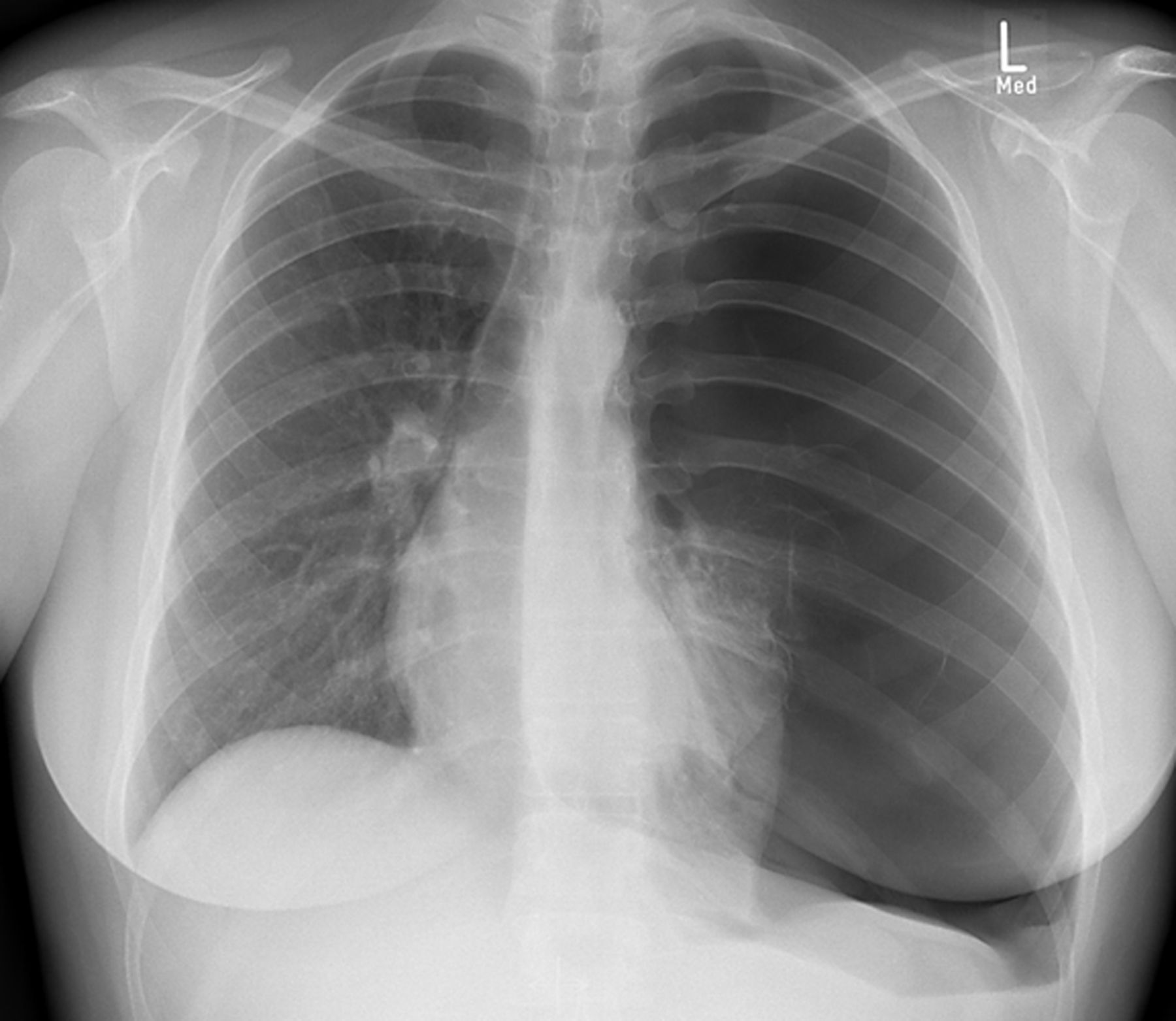
Main menu

  • Home
  • Current issue
  • Past issues
  • Authors/reviewers
    • Instructions for authors
    • Submit a manuscript
    • Peer reviewer login
  • Journal club
  • Alerts
  • Subscriptions
  • ERS Publications
    • European Respiratory Journal
    • ERJ Open Research
    • European Respiratory Review
    • Breathe
    • ERS Books
    • ERS publications home

User menu

  • Log in
  • Subscribe
  • Contact Us
  • My Cart

Search

  • Advanced search
  • ERS Publications
    • European Respiratory Journal
    • ERJ Open Research
    • European Respiratory Review
    • Breathe
    • ERS Books
    • ERS publications home

Login

European Respiratory Society

Advanced Search

  • Home
  • Current issue
  • Past issues
  • Authors/reviewers
    • Instructions for authors
    • Submit a manuscript
    • Peer reviewer login
  • Journal club
  • Alerts
  • Subscriptions

A persistent pneumothorax? 5-year follow-up after diagnosis of Swyer–James–MacLeod syndrome

Amanda Tufman, Hauke Winter, Kathrin Kahnert, Simone Reu, Ullrich Müller-Lisse, Rudolf Maria Huber
Breathe 2016 12: e55-E58; DOI: 10.1183/20734735.000316
Amanda Tufman
1Division of Respiratory Medicine and Thoracic Oncology, Dept of Internal Medicine V, Thoracic Oncology Centre Munich, Ludwig-Maximilians University, Munich, Germany
2Member of the German Center for Lung Research (DZL), Comprehensive Pneumology Center Munich, Munich, Germany
  • Find this author on Google Scholar
  • Find this author on PubMed
  • Search for this author on this site
  • For correspondence: amanda.tufman@med.uni-muenchen.de
Hauke Winter
2Member of the German Center for Lung Research (DZL), Comprehensive Pneumology Center Munich, Munich, Germany
3Dept of Thoracic Surgery, Thoracic Oncology Centre, Munich Ludwig Maximilians University, Munich, Germany
  • Find this author on Google Scholar
  • Find this author on PubMed
  • Search for this author on this site
Kathrin Kahnert
1Division of Respiratory Medicine and Thoracic Oncology, Dept of Internal Medicine V, Thoracic Oncology Centre Munich, Ludwig-Maximilians University, Munich, Germany
2Member of the German Center for Lung Research (DZL), Comprehensive Pneumology Center Munich, Munich, Germany
  • Find this author on Google Scholar
  • Find this author on PubMed
  • Search for this author on this site
Simone Reu
2Member of the German Center for Lung Research (DZL), Comprehensive Pneumology Center Munich, Munich, Germany
4Pathological Institute, Ludwig Maximilians University, Munich, Germany
  • Find this author on Google Scholar
  • Find this author on PubMed
  • Search for this author on this site
Ullrich Müller-Lisse
2Member of the German Center for Lung Research (DZL), Comprehensive Pneumology Center Munich, Munich, Germany
5Department of Radiology, Thoracic Oncology Centre Munich Ludwig Maximilians University, Munich, Germany
  • Find this author on Google Scholar
  • Find this author on PubMed
  • Search for this author on this site
Rudolf Maria Huber
1Division of Respiratory Medicine and Thoracic Oncology, Dept of Internal Medicine V, Thoracic Oncology Centre Munich, Ludwig-Maximilians University, Munich, Germany
2Member of the German Center for Lung Research (DZL), Comprehensive Pneumology Center Munich, Munich, Germany
  • Find this author on Google Scholar
  • Find this author on PubMed
  • Search for this author on this site
  • Article
  • Figures & Data
  • Info & Metrics
  • PDF
Loading

Figures

  • Additional Files
  • Figure 1
    • Download figure
    • Open in new tab
    • Download powerpoint
    Figure 1

    Chest radiograph upon presentation to the emergency department.

  • Figure 2
    • Download figure
    • Open in new tab
    • Download powerpoint
    Figure 2

    Chest radiography with partial re-expansion of the lung following chest tube insertion.

  • Figure 3
    • Download figure
    • Open in new tab
    • Download powerpoint
    Figure 3

    a) Coronal reconstruction of chest CT angiography images showing marked hypodensity of the left upper lobe. b) Coronal reconstruction of maximum intensity projection chest CT angiography images showing vascular hypoplasia of the left lung.
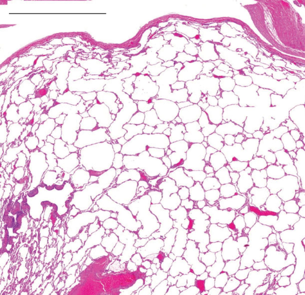
  • Figure 4
    • Download figure
    • Open in new tab
    • Download powerpoint
    Figure 4

    Emphysematous changes with hyperinflation in the resected lung tissue stained with haematoxylin and eosin. Scale bar=2 mm.

Additional Files

  • Figures
  • Disclosures

    Files in this Data Supplement:

    • U. Mueller-Lisse
PreviousNext
Back to top
Vol 12 Issue 2 Table of Contents
Breathe: 12 (2)
  • Table of Contents
  • Index by author
Email

Thank you for your interest in spreading the word on European Respiratory Society .

NOTE: We only request your email address so that the person you are recommending the page to knows that you wanted them to see it, and that it is not junk mail. We do not capture any email address.

Enter multiple addresses on separate lines or separate them with commas.
A persistent pneumothorax? 5-year follow-up after diagnosis of Swyer–James–MacLeod syndrome
(Your Name) has sent you a message from European Respiratory Society
(Your Name) thought you would like to see the European Respiratory Society web site.
CAPTCHA
This question is for testing whether or not you are a human visitor and to prevent automated spam submissions.
Print
Citation Tools
A persistent pneumothorax? 5-year follow-up after diagnosis of Swyer–James–MacLeod syndrome
Amanda Tufman, Hauke Winter, Kathrin Kahnert, Simone Reu, Ullrich Müller-Lisse, Rudolf Maria Huber
Breathe Jun 2016, 12 (2) e55-E58; DOI: 10.1183/20734735.000316

Citation Manager Formats

  • BibTeX
  • Bookends
  • EasyBib
  • EndNote (tagged)
  • EndNote 8 (xml)
  • Medlars
  • Mendeley
  • Papers
  • RefWorks Tagged
  • Ref Manager
  • RIS
  • Zotero

Share
A persistent pneumothorax? 5-year follow-up after diagnosis of Swyer–James–MacLeod syndrome
Amanda Tufman, Hauke Winter, Kathrin Kahnert, Simone Reu, Ullrich Müller-Lisse, Rudolf Maria Huber
Breathe Jun 2016, 12 (2) e55-E58; DOI: 10.1183/20734735.000316
Reddit logo Technorati logo Twitter logo Connotea logo Facebook logo Mendeley logo
Full Text (PDF)

Jump To

  • Article
    • Abstract
    • Discussion
    • Footnotes
    • References
  • Figures & Data
  • Info & Metrics
  • PDF

Subjects

  • Lung imaging
  • Respiratory clinical practice
  • Tweet Widget
  • Facebook Like
  • Google Plus One

More in this TOC Section

Expert opinion

  • Lung cancer screening by volume CT
  • In pursuit of the primary
  • A rare complication in a case of nonsmall cell lung carcinoma
Show more Expert opinion

Case report

  • Cutaneous lesions in a COVID-19 patient leading to a surprising diagnosis
  • Chronic cough and inflammatory bowel disease
  • Bilateral mediastinal lymphadenopathy with cough and shortness of breath
Show more Case report

Related Articles

Navigate

  • Home
  • Current issue
  • Archive

About Breathe

  • Journal information
  • Editorial board
  • Press
  • Permissions and reprints
  • Advertising

The European Respiratory Society

  • Society home
  • myERS
  • Privacy policy
  • Accessibility

ERS publications

  • European Respiratory Journal
  • ERJ Open Research
  • European Respiratory Review
  • Breathe
  • ERS books online
  • ERS Bookshop

Help

  • Feedback

For authors

  • Intructions for authors
  • Publication ethics and malpractice
  • Submit a manuscript

For readers

  • Alerts
  • Subjects
  • RSS

Subscriptions

  • Accessing the ERS publications

Contact us

European Respiratory Society
442 Glossop Road
Sheffield S10 2PX
United Kingdom
Tel: +44 114 2672860
Email: journals@ersnet.org

ISSN

Print ISSN: 1810-6838
Online ISSN: 2073-4735

Copyright © 2023 by the European Respiratory Society