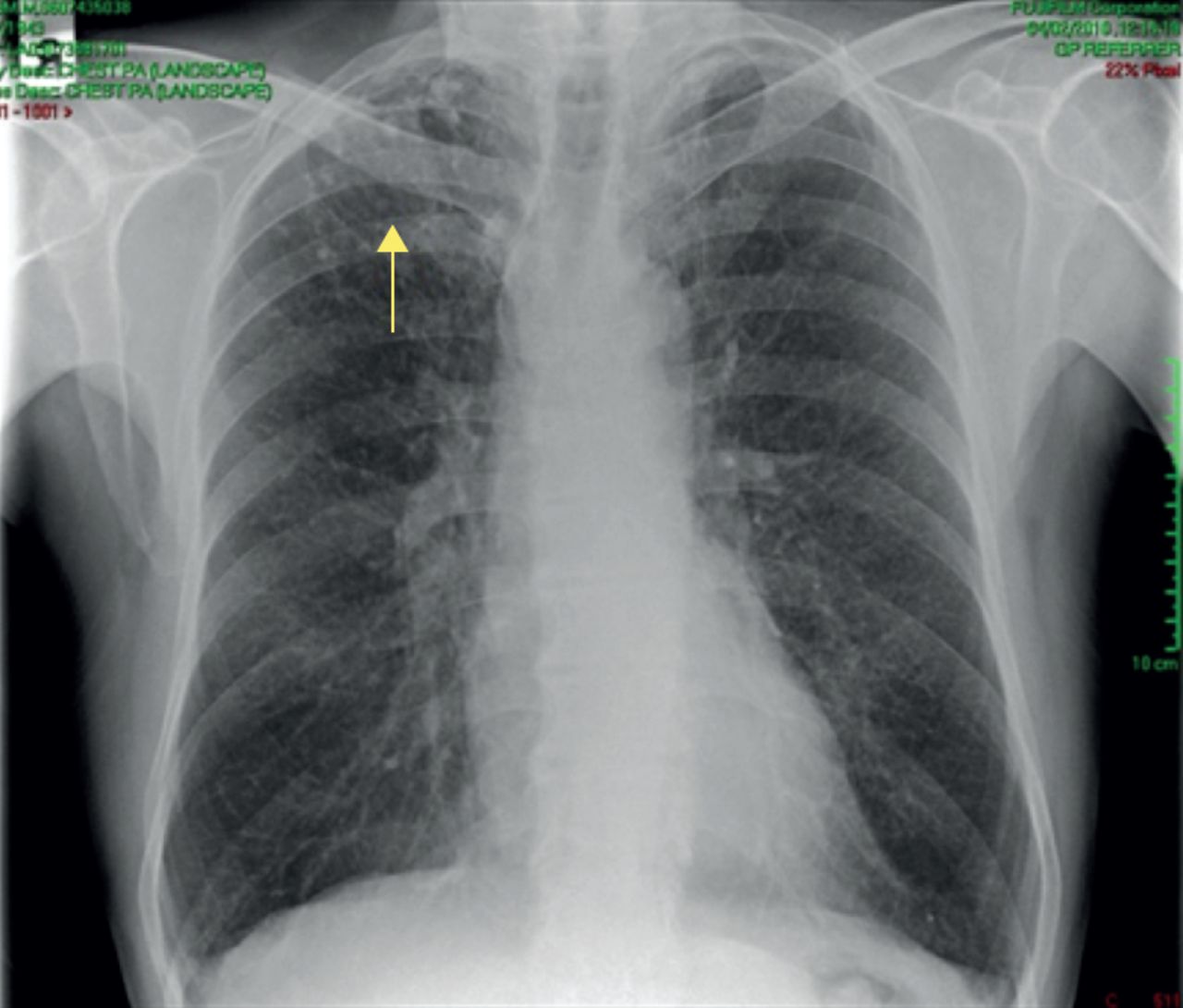
Main menu

  • Home
  • Current issue
  • Past issues
  • Authors/reviewers
    • Instructions for authors
    • Submit a manuscript
    • Peer reviewer login
  • Journal club
  • Alerts
  • Subscriptions
  • ERS Publications
    • European Respiratory Journal
    • ERJ Open Research
    • European Respiratory Review
    • Breathe
    • ERS Books
    • ERS publications home

User menu

  • Log in
  • Subscribe
  • Contact Us
  • My Cart

Search

  • Advanced search
  • ERS Publications
    • European Respiratory Journal
    • ERJ Open Research
    • European Respiratory Review
    • Breathe
    • ERS Books
    • ERS publications home

Login

European Respiratory Society

Advanced Search

  • Home
  • Current issue
  • Past issues
  • Authors/reviewers
    • Instructions for authors
    • Submit a manuscript
    • Peer reviewer login
  • Journal club
  • Alerts
  • Subscriptions

A suspected bronchial carcinoma

Sofia G. Korsavva, Sudipta Roy
Breathe 2017 13: e114-e120; DOI: 10.1183/20734735.010317
Sofia G. Korsavva
Respiratory Medicine, Hairmyres District Hospital, Glasgow, UK
  • Find this author on Google Scholar
  • Find this author on PubMed
  • Search for this author on this site
  • For correspondence: sophiadr@live.com
Sudipta Roy
Respiratory Medicine, Hairmyres District Hospital, Glasgow, UK
  • Find this author on Google Scholar
  • Find this author on PubMed
  • Search for this author on this site
  • Article
  • Figures & Data
  • Info & Metrics
  • PDF
Loading

Figures

  • Figure 1
    • Download figure
    • Open in new tab
    • Download powerpoint
    Figure 1

    Baseline chest radiograph, February 2010.

  • Figure 2
    • Download figure
    • Open in new tab
    • Download powerpoint
    Figure 2

    Repeat chest radiograph in 2011.
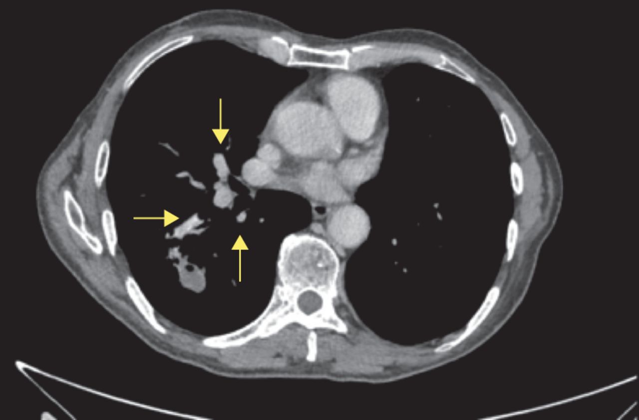
  • Figure 3
    • Download figure
    • Open in new tab
    • Download powerpoint
    Figure 3

    CT scan, August 2011.

  • Figure 4
    • Download figure
    • Open in new tab
    • Download powerpoint
    Figure 4

    Initial PET–CT scan showing “hot spots”.

  • Figure 5
    • Download figure
    • Open in new tab
    • Download powerpoint
    Figure 5

    Post-lobectomy chest radiograph.

  • Figure 6
    • Download figure
    • Open in new tab
    • Download powerpoint
    Figure 6

    a) Iron stain showing welder’s lung and various lung segments consistent with welder’s lung changes: fibrosis with carbon and iron pigment. b) Lung tissue with scarring, abundant histiocytes and multinucleated giant cells containing pigment both iron and carbon. The alveoli contain numerous macrophages and chronic inflammatory cells. At higher magnification, there is dystrophic calcification and ossification. c and d) Lymph nodes with a lot of pigment deposition but no evidence of malignancy.

  • Figure 7
    • Download figure
    • Open in new tab
    • Download powerpoint
    Figure 7

    CT scan, September 2012. Several new lung masses can be seen. There is a cavitating soft tissue mass measuring 2.5×1.9 cm within the right lower lobe with adjacent consolidation and inflammatory changes. There is a further mass within the left upper lobe measuring 1.3 cm. No other sizeable intrapulmonary mass lesions are seen. A precarinal node measures 9 mm in the short axis. These are highly suggestive of malignancy.

  • Figure 8
    • Download figure
    • Open in new tab
    • Download powerpoint
    Figure 8

    Follow up PET–CT scan.
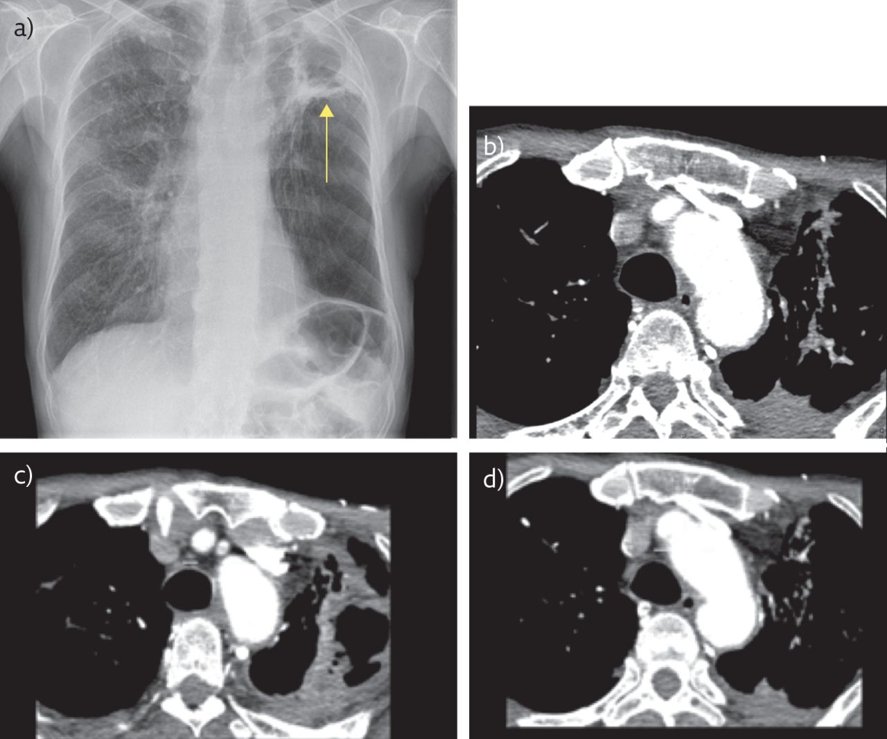
  • Figure 9
    • Download figure
    • Open in new tab
    • Download powerpoint
    Figure 9

    a) Chest radiograph (2016) of the patient showing areas of old TB disease (arrow). b–d) Thoracic CT thorax snapshots showing areas of marked fibrosis.
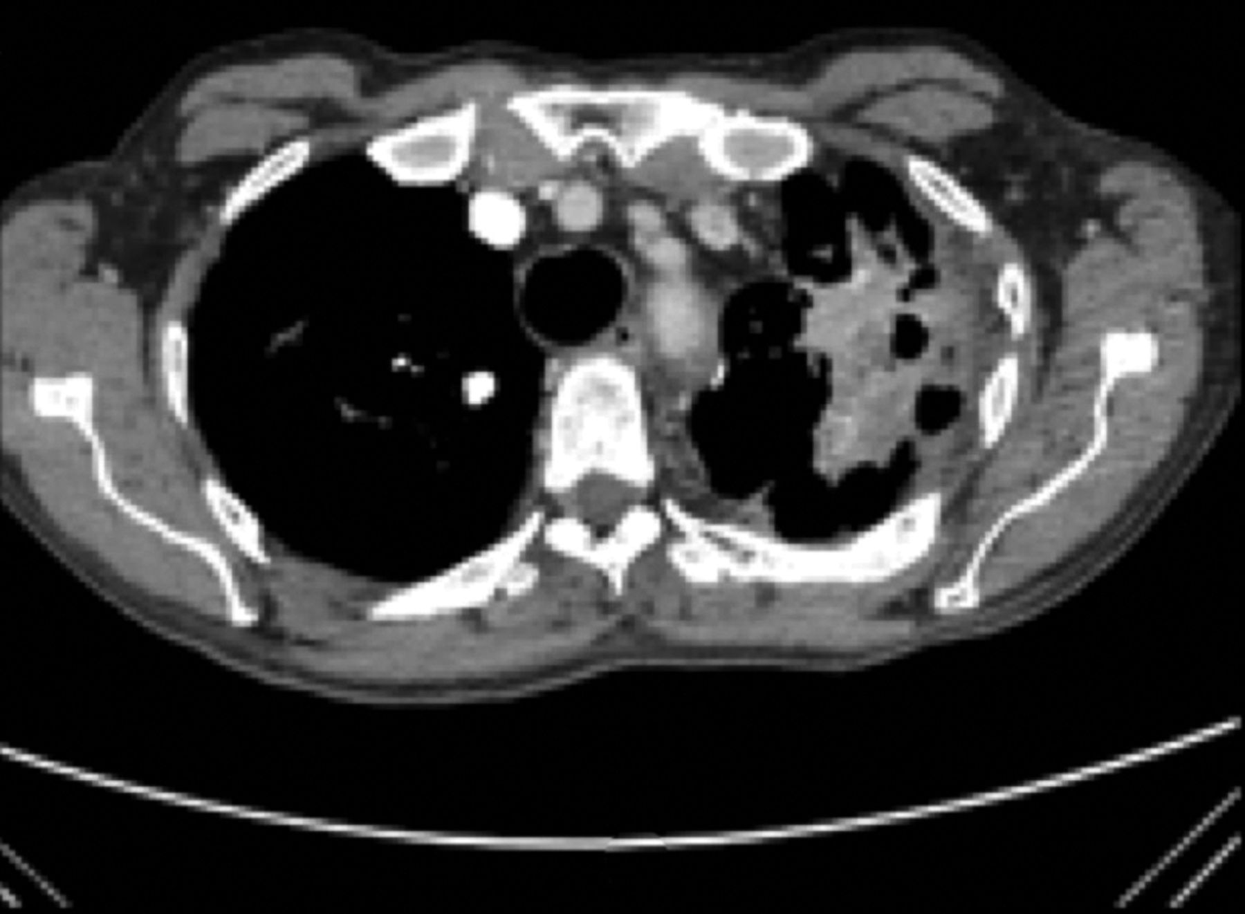
  • Figure 10
    • Download figure
    • Open in new tab
    • Download powerpoint
    Figure 10

    CT scan, June 2015. There is a thick-walled cavity in the left apex at the site of the previous bulla that incorporates the previously noted 1.5-cm soft tissue nodule.

  • Figure 11
    • Download figure
    • Open in new tab
    • Download powerpoint
    Figure 11

    CT scan with contrast, 2016. A further increase in the cavitation in the left upper zone with marked related emphysema in the residual left lower lobe can be seen.

  • Figure 12
    • Download figure
    • Open in new tab
    • Download powerpoint
    Figure 12

    CT scan, June 2105. A cavitating lesion can be seen in the right lobe.

  • Figure 13
    • Download figure
    • Open in new tab
    • Download powerpoint
    Figure 13

    CT scan, October 2016. The lesion has now become a spiculated nodule.

  • Figure 14
    • Download figure
    • Open in new tab
    • Download powerpoint
    Figure 14

    Latest chest radiograph.

PreviousNext
Back to top
Vol 13 Issue 4 Table of Contents
Breathe: 13 (4)
  • Table of Contents
  • Index by author
Email

Thank you for your interest in spreading the word on European Respiratory Society .

NOTE: We only request your email address so that the person you are recommending the page to knows that you wanted them to see it, and that it is not junk mail. We do not capture any email address.

Enter multiple addresses on separate lines or separate them with commas.
A suspected bronchial carcinoma
(Your Name) has sent you a message from European Respiratory Society
(Your Name) thought you would like to see the European Respiratory Society web site.
CAPTCHA
This question is for testing whether or not you are a human visitor and to prevent automated spam submissions.
Print
Citation Tools
A suspected bronchial carcinoma
Sofia G. Korsavva, Sudipta Roy
Breathe Dec 2017, 13 (4) e114-e120; DOI: 10.1183/20734735.010317

Citation Manager Formats

  • BibTeX
  • Bookends
  • EasyBib
  • EndNote (tagged)
  • EndNote 8 (xml)
  • Medlars
  • Mendeley
  • Papers
  • RefWorks Tagged
  • Ref Manager
  • RIS
  • Zotero

Share
A suspected bronchial carcinoma
Sofia G. Korsavva, Sudipta Roy
Breathe Dec 2017, 13 (4) e114-e120; DOI: 10.1183/20734735.010317
Reddit logo Technorati logo Twitter logo Connotea logo Facebook logo Mendeley logo
Full Text (PDF)

Jump To

  • Article
    • Abstract
    • Examination
    • Investigations
    • Discussion
    • Conclusion
    • Acknowledgements
    • Footnotes
    • References
  • Figures & Data
  • Info & Metrics
  • PDF

Subjects

  • Respiratory clinical practice
  • Tweet Widget
  • Facebook Like
  • Google Plus One

More in this TOC Section

Expert opinion

  • Lung cancer screening by volume CT
  • In pursuit of the primary
  • Contribution of causal factors to disease burden
Show more Expert opinion

Case report

  • Chronic cough and inflammatory bowel disease
  • Cutaneous lesions in a COVID-19 patient leading to a surprising diagnosis
  • Bilateral mediastinal lymphadenopathy with cough and shortness of breath
Show more Case report

Related Articles

Navigate

  • Home
  • Current issue
  • Archive

About Breathe

  • Journal information
  • Editorial board
  • Press
  • Permissions and reprints
  • Advertising

The European Respiratory Society

  • Society home
  • myERS
  • Privacy policy
  • Accessibility

ERS publications

  • European Respiratory Journal
  • ERJ Open Research
  • European Respiratory Review
  • Breathe
  • ERS books online
  • ERS Bookshop

Help

  • Feedback

For authors

  • Intructions for authors
  • Publication ethics and malpractice
  • Submit a manuscript

For readers

  • Alerts
  • Subjects
  • RSS

Subscriptions

  • Accessing the ERS publications

Contact us

European Respiratory Society
442 Glossop Road
Sheffield S10 2PX
United Kingdom
Tel: +44 114 2672860
Email: journals@ersnet.org

ISSN

Print ISSN: 1810-6838
Online ISSN: 2073-4735

Copyright © 2023 by the European Respiratory Society