


Figures
- Figure 1
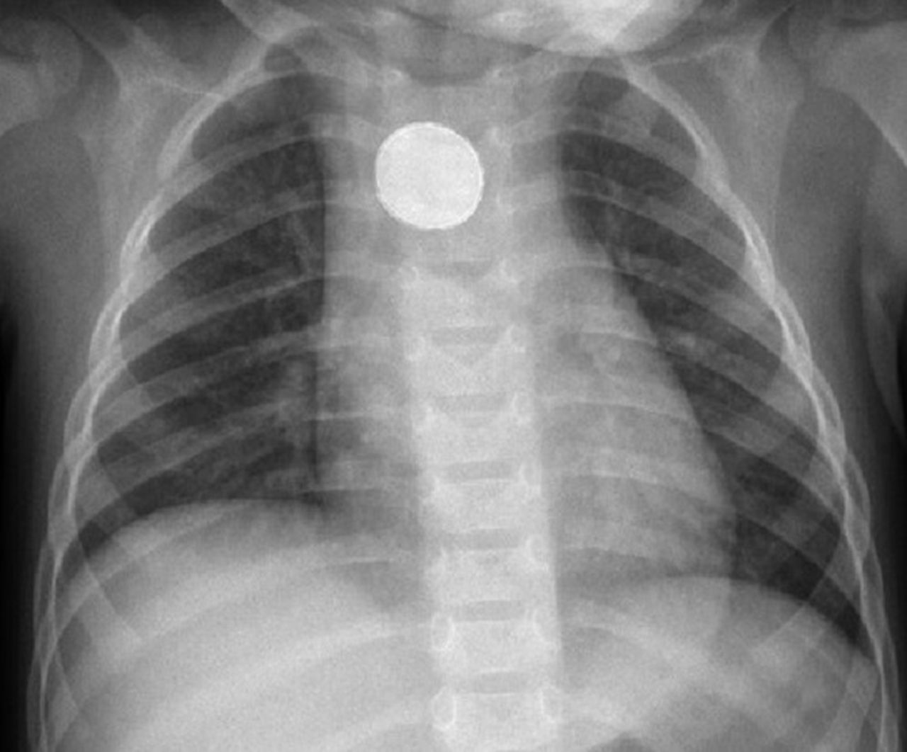
Chest radiogram of a 16-month-old child with history of stridor for 3 months.
- Figure 2
Endoscopic evaluation of oesophagus in a 16-month-old child with history of stridor for 3 months. a) Foreign body at the level of the second oesophageal constriction. b) Foreign body (lithium battery) after removal from the oesophagus.


Tables
- Table 1
Characteristics and additional diagnostic techniques to differentiate the causes of stridor in children
Characteristics Additional diagnostic techniques Acute Foreign body aspiration or ingestion [5–7] Peak age 1–3 years
Sudden-onset coughing and choking that might be followed by symptom-free period, and thus be misinterpreted as resolution
Potentially life threateningChest radiography
CT when suspected that negative result might avert bronchoscopy
BronchoscopyAnaphylaxis Potentially life threatening
Possible additional symptoms (skin and/or gastrointestinal)Detail history of the episode Infection Bacterial tracheitis [7] Any age; most commonly, first 6 years
ARVI-like prodromal period
Croup-like symptoms that do not respond to standard croup therapyDirect laryngoscopy and/or bronchoscopy gives a definitive diagnosis but is not routinely performed
Specimens for aetiological diagnosis during endoscopy immediately after intubation; older patients might provide sputumEpiglottitis [7] Decreased incidence and increased age at presentation (previously 3 years, now 6–12 years) since Hib vaccine was introduced
Sudden onset, rapid progression
Hallmark: three D's (dysphagia, drooling and distress), fever, toxic appearance, hoarse voice, stridor, pharyngitis
Various degrees of severity
Young children: respiratory distress, anxiety, “tripod”/ “sniffing” posture, drooling; cough not characteristic
Older children: might just have severely sore throatOften clinical diagnosis
Direct laryngoscopy (swollen epiglottitis)
Lateral radiography of the neck, looking for the “thumb sign”
Laboratory tests and microbiology only if the airways are safe
Need for a very cautious examination is warranted in theatre with experienced anaesthetist and an ENT specialist capable of performing an emergency airway procedureDiphtheria [7] Presenting symptoms: malaise, sore throat, fever (low grade), cervical lymphadenopathy
Mild pharyngeal erythema → isolated exudate (grey, white) → pseudomembrane (at least one third of cases); pseudomembrane can extend to lower parts of respiratory system
Laryngeal diphtheria (pseudomembrane covers larynx) might be isolated (cough, hoarseness) or a part of malignant diphtheria (stridor, respiratory insufficiency)
Systemic manifestations: myocarditis, neuropathiesCulture of Corynebacterium diphtheriae from respiratory tract
Toxin detection
Laryngoscopy: pseudomembraneAirway burns Thermal epiglottitis and upper airway burns [8] Clinical presentation similar to that of infectious epiglottitis; might not correlate with severity, especially in younger children
With/without cutaneous burn injury
Risk of rapid airway obstruction (because of developing oedema)Direct laryngoscopy
BronchoscopyCaustic burns [9] More common 1–3 years of age
Upper airway involvement: hoarseness, stridor, nasal flaring, retractions
Other symptoms: food refusal, drooling, dysphagia (oropharyngeal/oesophageal injury)
Symptoms might not correlate with severity, especially in younger children
May be misdiagnosed as anaphylaxisDirect laryngoscopy
BronchoscopySubacute Retropharyngeal abscess [7, 10, 11] Peaks at 2–4 years of age
Often after upper airway infection (tonsillitis, pharyngitis, lymphadenitis)
Early stage: symptoms indistinguishable from uncomplicated pharyngitis
Later stage: dysphagia, odynophagia, drooling, torticollis, neck pain, dysphonia, respiratory distress, stridor, trismus, fever, chest pain
Symptoms might be similar to that of epiglottitis but progress slowerLateral neck radiograph (might be false positive if the child is crying)
CT scan with intravenous contrastPeritonsillar abscess [7, 12] More often in adolescents
Severe sore throat (mainly unilateral), fever, muffled voice, trismus, droolingPus drainage from abscess confirms diagnosis
Laboratory tests not necessary
Imaging studies not routinely performed; might help differentiate peritonsillar abscess from cellulitis (intraoral or submandibular US), deep space neck infection (CT scan with contrast) and epiglottitis (direct laryngoscopy, lateral neck radiograph)Chronic/recurrent Congenital Laryngomalacia [5, 7, 13] Usually begins at neonatal period: 4–5 weeks, peaks at 4–8 months; may resolve by 12–18 months
Inspiratory “wet” low-pitch stridor; hoarseness is atypical
May worsen in the supine and improve in the prone position
Worsens during respiratory infections
Mild to moderate: louder when sleeping and feeding; may disappear when crying
Severe: louder when crying.
Severe: associated with other problems (sleep disordered breathing, failure to thrive etc.)
Higher incidence of gastro-oesophageal refluxFlexible laryngoscopy if associated problems are noted (failure to thrive, apnoea, significant/progressive stridor, etc.)
Sleep endoscopy: suspicion of state dependent laryngomalacia (during sleep)Tracheomalacia [5, 14, 15] Usually manifests from 2–3 months of age
More common in children with oesophageal atresia
Barking or brassy cough, stridor
Moderate: more frequent lower airway infections
Severe: upper respiratory tract obstruction, cyanosis, apnoeic spells
Symptoms might become more evident with activities (crying, eating)Dynamic airway endoscopy: diagnostic tool of choice
CT scan: end-expiratory and end-inspiratory images (endotracheal intubation needed in young kids)
Free-breathing cine CT scan (can be used in young children, does not require breathing manoeuvres cooperation)
Barium oesophagography (evaluating tracheal compression by oesophagus or other structures)Vocal cord paralysis [5, 7, 16] Onset of symptoms: birth to 5 years
Bilateral (birth trauma, neurological, unknown reason): stridor, respiratory insufficiency, cyanosisFlexible fibreoptic nasopharyngolaryngoscopy
Direct laryngoscopy
Laryngeal ultrasoundVascular ring [17] Great clinical variability from critical airway obstruction to asymptomatic (incomplete vascular ring)
Stridor (usually louder during expiration), wheezing, cough, respiratory distress, respiratory infections
Digestive system complaints: dysphagia, feeding difficulty, vomiting (complete vascular ring)
Associated anomalies: congenital heart disease, tracheo-oesophageal fistula, cleft lip/palate, subglottic stenosis, genetic or malformation syndromesAnterior, posterior, lateral chest radiograph (compressed trachea, anterior bowing of the trachea)
CT scan or MRA
Echocardiography
Bronchoscopy (not routinely performed)
Barium swallow (not routinely performed)Bronchogenic cyst [14] Usually presents in adolescence with recurrent cough, wheezing (might simulate asthma), pneumonia
Starting in infancy: respiratory distress, cyanosis, feeding difficultyChest radiograph
CT scan, MRILaryngeal malformations [13] Cyst (vallecular, saccular), laryngocele, stenosis, cleft usually present in infancy/early childhood
Stridor, wheezing, noisy breathing, hoarseness, aspiration, recurrent respiratory infections, feeding difficulty, failure to thriveEndoscopy Infantile haemangiomas [5, 7, 13, 18] Symptoms typically start at 1–3 months and resolve by 5–12 years of age
Presentation similar to that of subglottic stenosis; recurrent croup, biphasic stridor (may progress to respiratory distress)
Initially might be misdiagnosed as croup; response to standard croup therapy is transient
Might be associated with other haemangiomas, especially in the “beard” distributionEndoscopy
Radiograph of the neck: asymmetric narrowing of the subglottis
CT scan with contrast: delineatingSubglottic stenosis [5, 7, 13, 19] Biphasic stridor, recurrent episodes of croup and barking cough
Typically improves with timeEndoscopy Acquired VCD or paradoxical vocal fold motion [20, 21] Transient improper adduction of the true vocal folds (inspiration and/or expiration)
Great clinical variability; may also mimic other diseases (e.g. asthma attack), frequently misdiagnosed
Various triggers: exercise, stress, irritants, infections, etc.
Stridor, globus sensation, difficulty swallowing, chest tightness, aphonia/dysphonia, sensation of choking that can lead to stress, anxiety, panic
Usually self limitingVCDQ (symptom monitoring)
Pittsburgh VCD index (differential diagnosis with asthma)
Direct flexible laryngoscopy (if possible, after bronchoprovocation challenge) is the gold standard
Pulmonary function testing (possible changes in inspiratory loop)
Impulse oscillometryRecurrent respiratory papillomatosis [22, 23] Juvenile (usually more aggressive; most commonly 2–4 years of age) or adult (diagnosis after 12 years of age) onset
Hoarseness: usually the presenting symptom, followed by stridor.
Less often: failure to thrive, chronic cough, dysphagia, dyspnoea, acute respiratory distress, recurrent pneumonia.
Often misdiagnosed as croup, asthma, allergies, bronchitis, vocal nodules. The diagnosis usually made 1 year after the onset of symptomsLaryngoscopy Vocal cord paralysis Unilateral (usually iatrogenic): hoarse voice, crying affections; risk for aspiration Flexible fibreoptic nasopharyngolaryngoscopy
Direct laryngoscopy
Laryngeal ultrasoundSubglottic stenosis [24] Symptoms similar but less severe to that of congenital subglottic stenosis Endoscopy Hypocalcaemic laryngeal spasm [25] Children with vitamin D deficiency and rickets (mostly); metabolic/endocrine disorders that result in hypocalcaemia
Stridor: chronic intermittent or acute and severe; rarely as presenting symptom
Other symptoms due to hypocalcaemia: muscle contractions, anticonvulsant-resistant seizures; in neonates: apnoea, lethargy, poor feeding, abdominal distension, tachycardia, vomitingChvostek or Trousseau sign
Blood tests: electrolytes, alkaline phosphatase, phosphate, magnesium, PTH, vitamin D metabolites, liver function tests
ECG: prolonged QTc
Urine tests: pH, calcium, magnesium, phosphate, creatinine
Urine calcium/creatinine ratioTumour [26] Tumors that compress the airways may present with expiratory stridor, shortness of breath, cough, hoarse voice Chest radiograph: mediastinal masses are commonly discovered on routine test GORD [27, 28] Nocturnal stridor and cough are atypical manifestations of GORD
Other symptoms
Infants: feeding refusal, poor weight gain, haematemesis, anaemia, respiratory symptoms
Preschool: intermittent regurgitation, respiratory symptoms, decreased food intake and poor weight gain; Sandifer syndrome
School-aged children and adolescents: postprandial cough, chronic cough, hoarseness, dysphagia, globus sensation, bitter taste in mouth, heartburn, nauseaEmpiric treatment
24-h pH monitoring or impedance monitoring
Endoscopy and histologyCT: computed tomography; ARVI: acute respiratory viral infection; Hib: Haemophilus influenzae type b; ENT: ear, nose and throat; US: ultrasonography; MRA: magnetic resonance angiography; MRI: magnetic resonance imaging; VCD: vocal cord dysfunction; VCDQ: Vocal Cord Dysfunction Questionnaire; PTH: parathyroid hormone; QTc: QT interval; GORD: gastro-oesophageal reflux disease.


