


Figures
- Figure 1
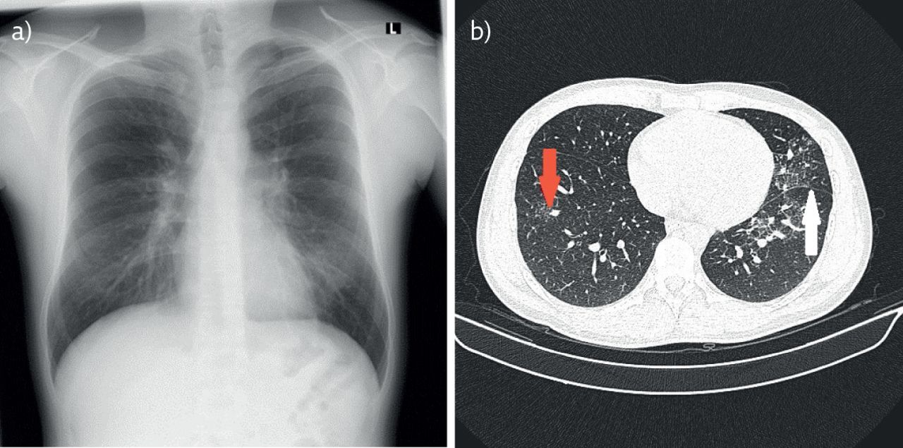
a) Chest radiograph showing infiltrate in the left lung. b) HRCT of the thorax, red arrow pointing to the micronodular pattern and white arrow pointing to septal thickening.
- Figure 2
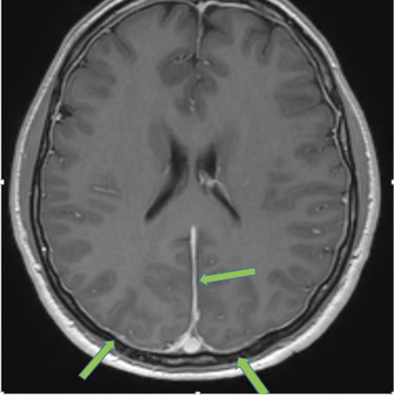
Thickening and imbibition of dura on brain MRI.
- Figure 3
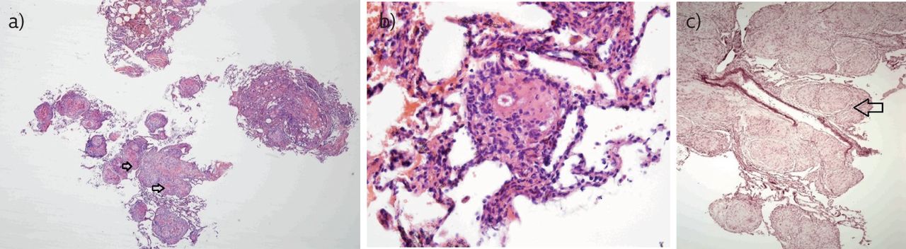
a–c) Transbronchial lung biopsy samples.
Vol 17 Issue 4
Table of Contents

A young man with transitory hemiparesis and lung infiltrates


