


Abstract
This case describes an uncommon presentation of type 2 respiratory failure secondary to left hemidiaphragmatic paralysis. Emphasis is on the multitude of possible causes of hemidiaphragmatic paralysis and how to manage such a presentation. https://bit.ly/3Mcd2XI
A 73-year-old man presented to the emergency department with worsening orthopnoea and exertional dyspnoea. He denied any other cardiorespiratory symptoms. The patient had a body mass index (BMI) of 23.3 kg·m−2 (height 163 cm, weight 62 kg). Lung function tests 3 months before admission showed a ratio of forced expiratory volume in 1 s (FEV1) to forced vital capacity (FVC) of 88%. He had a past medical history of hypothyroidism, for which he was on levothyroxine 50 μg daily, and type 2 diabetes mellitus, for which he was on metformin 500 mg twice daily. Past surgical history included a gastrectomy 3 years prior to admission performed for gastric cancer, which was subsequently complicated by a subphrenic abscess, pancreatitis and residual pancreatic insufficiency. The patient was a lifelong non-smoker, worked in construction, reared birds as a hobby, and lived in an urban area. He was not on any other medication.
On examination he was noted to be tachypnoeic with a respiratory rate of 40 breaths·min−1. A flap of his palms was noted but his chest revealed normal breath sounds on auscultation. Neurological examination elicited few fasciculations in the right triceps and left quadriceps, decreased reflexes throughout both upper limbs and absent ankle jerk bilaterally. He had no sensory deficits and no evident upper motor neurone signs suggesting major neurological diseases of the brain or spinal cord.
The patient's physiological observations were stable with oxygen saturation measured by pulse oximetry (SpO2) of 95% on room air. Routine blood tests were unremarkable with a normal complete blood count, normal renal profile, and normal C-reactive protein. Thyroid function tests showed an elevated thyroid-stimulating hormone level of 3.994 μIU·mL−1 with a normal thyroxine level. The levothyroxine dose was subsequently increased from 50 μg to 75 μg daily. An ECG was unremarkable.
Task 1
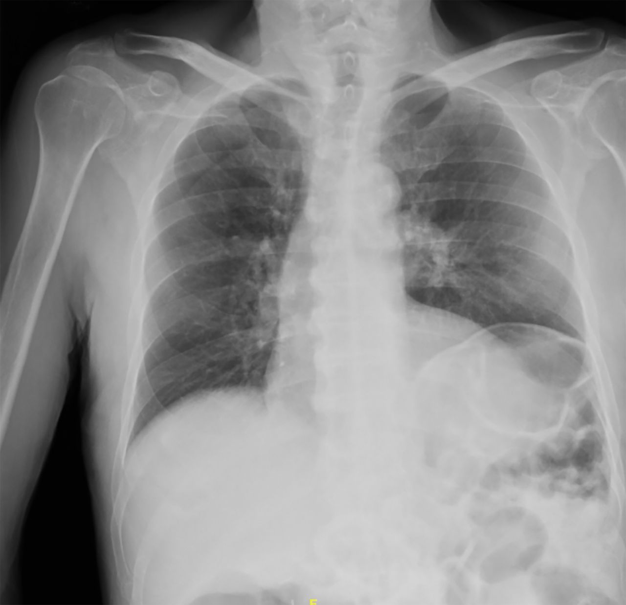
What can you note on this chest radiograph taken on admission (figure 1)?

Chest radiograph taken on admission.
Answer 1
This chest radiograph is taken anteroposteriorly. There is an elevated left hemidiaphragm and surgical clips in the right upper quadrant. No other abnormalities can be noted.
N-terminal pro-brain natriuretic peptide (NT-proBNP) was found to be mildly elevated at 458 pg·mL−1, for which the patient was started on bumetanide 0.5 mg daily. An echocardiogram noted normal left ventricular dimensions, mildly impaired global left ventricular systolic function with an ejection fraction of 45%, normal right ventricular function and no valvular lesions. The patient was empirically started on controlled low-dose oxygen via a 24% Venturi mask at 2 L·min−1, on an at-request basis when complaining of shortness of breath. Arterial blood gases at rest on 24% oxygen showed pH 7.33, arterial carbon dioxide tension (PaCO2) 81 mmHg (10.8 kPa), arterial oxygen tension (PaO2) 93 mmHg (12.4 kPa), SpO2 96%, lactate 0.8 mmol·L−1 and HCO3− 43 mmol·μL−1.
Task 2
How would you interpret the arterial blood gas results?
a) Respiratory acidosis with partial metabolic compensation
b) Respiratory acidosis without metabolic compensation
c) Metabolic acidosis with partial respiratory compensation
d) Metabolic acidosis without respiratory compensation
e) Respiratory alkalosis
f) Metabolic alkalosis
Answer 2
a. Respiratory acidosis with partial metabolic compensation.
Task 3
What is the next best step in the management of this patient's type 2 respiratory failure?
a) Nasal prongs at 2 L·min−1
b) Normal face mask at 7 L·min−1
c) Non-rebreather mask at 10–15 L·min−1
d) Non-invasive ventilation
e) Invasive mechanical ventilation
Answer 3
d. Non-invasive ventilation.
The patient was admitted to a medical ward and was started on non-invasive ventilation (NIV). NIV settings were titrated upwards to an inspiratory positive airway pressure (IPAP) of 17 cmH2O with an expiratory positive airway pressure (EPAP) of 5 cmH2O, from an initial setting of 12 cmH2O IPAP. Attempts at weaning by alternating 2 h of NIV with rest periods of 2 h during the day at 24% oxygen throughout his stay were initially unsuccessful, with rises of PaCO2 to 102 mmHg (13.6 kPa) and drops in pH to 7.298.
Computed tomography (CT) and magnetic resonance imaging (MRI) of the brain only showed small vessel ischaemic disease but were otherwise unremarkable. There was no evidence of cerebral infarction, haemorrhage, or space-occupying lesions such as subdural haematoma or tumour. Further evaluation of the elevated left hemidiaphragm was subsequently performed using CT imaging of the thorax, abdomen and pelvis. A high position of the left hemidiaphragm was noted but thoracic imaging was otherwise unremarkable. Abdominal images did not show an acute process that could be responsible for the weakness of the left hemidiaphragm.
A high-resolution CT scan of the lungs was unremarkable and confirmed the clinical impression that emphysema and interstitial lung disease due to hypersensitivity pneumonitis to birds were not present. The patient was referred for a fluoroscopic sniff test, which showed elevation of the left hemidiaphragm, decreased movement of the left hemidiaphragm but no paradoxical movement. Subsequently, interrogation of both hemidiaphragms was performed using thoracic ultrasonography. The right hemidiaphragm showed normal diaphragmatic excursion on a tidal breath and sniff manoeuvre but there was complete left hemidiaphragmatic paralysis (figure 2).

M-mode thoracic ultrasound showing no left hemidiaphragmatic excursion, indicative of left hemidiaphragmatic paralysis.
Pulmonary function testing was used to quantify the degree of diaphragmatic dysfunction, which showed FEV1 of 1.6 L (67% predicted), FVC of 1.84 L (58% pred), peak expiratory flow rate of 255 L·min−1 (61% pred) and FEV1/FVC ratio of 88% with no significant reversibility. FVC repeated in the supine position was 1.46 L (46% pred). Diffusing capacity of the lung for carbon monoxide (DLCO) was 11.7 mL·min−1·mmHg−1 (77% pred) and alveolar volume (VA) was 3.7 L, with DLCO/VA of 3.17 mL·min−1·mmHg−1·L−1 (89% pred).
Task 4
How would you interpret these pulmonary function tests?
a) Normal
b) Obstructive
c) Restrictive
d) Mixed
Answer 4
c. Restrictive.
Investigations were performed because of a possible myopathy, neuromuscular junction pathology or left phrenic neuropathy. Erythrocyte sedimentation rate, creatine kinase and aldolase were unremarkable. An auto-antibody screen for anti-mitochondrial antibodies, anti-smooth muscle antibodies, anti-gastric parietal cell antibodies and liver and kidney microsomal antibodies was negative. Anti-acetylcholine receptor antibodies and anti-muscle-specific kinase antibodies were also negative. Immunoglobulins G, M and A were within normal limits. HIV, hepatitis and syphilis serology were unremarkable. Serum protein electrophoresis did not identify a monoclonal band. Vitamin B12 and folate levels were within normal limits. Significant findings included an elevated lead level at 90 pg·dL−1 (normal level in adults is <25 μg·dL−1).
MRI of the cervical spine showed degenerative spine disease with a left mediolateral disc protrusion at the C3/C4 level (figure 3). The patient was also referred for nerve conduction studies, which showed chronic denervation/reinnervation changes, fasciculations in most of the muscles of the upper and lower extremities, as well as ongoing active denervation changes in the right tibialis anterior muscle, in keeping with a neurogenic process affecting mainly the motor nerves, which, however, did not fulfil El Escorial criteria for amyotrophic lateral sclerosis [1].

MRI of the spine. Axial T2 gradient echo sequence image at the level of C3/C4 shows a left mediolateral disc protrusion with impingement of the cord and the left C4 nerve root (dark area indicated by arrow).
On the fifth day after admission, the patient was eventually successfully weaned off continuous NIV, using progressively longer intervals without ventilation. He was provided with a portable NIV machine on pressure support mode with an IPAP of 16 cmH2O and an EPAP of 6 cmH2O to be used for 6–8 h at night. He was discharged 14 days after admission with significant resolution of symptoms and respiratory acidosis. He is being followed-up by a respiratory team and a neurology team. He stated that he is compliant with treatment and is still well with persistent resolution of symptoms, 6 months after discharge.
Discussion
The most likely cause of this patient's respiratory acidosis was left hemidiaphragmatic paralysis due to left phrenic neuropathy secondary to cervical spondylosis affecting the origin of the left phrenic nerve, as evidenced by the findings on MRI. Damage to the phrenic nerve could also have occurred following the previous gastric surgery or the complicating abscess or invasion by the tumour. The patient had a normal BMI, making obesity hypoventilation syndrome very unlikely, while his lung function tests were not diagnostic of COPD, and emphysema was not noted on the CT scan. The patient had some clinical evidence of diabetic peripheral neuropathy, with a possible motor neuropathy due to lead exposure. However, this would have affected both diaphragms equally. Notwithstanding, this could have contributed to the ventilation failure on top of the diaphragmatic weakness. The echocardiogram and the NT-proBNP suggested mild left ventricular failure, which is not usually a cause of respiratory acidosis, but rather of type 1 respiratory failure and only when severe.
We describe a case of an uncommon aetiology of type 2 respiratory failure secondary to left hemidiaphragmatic paralysis. Respiratory failure is conventionally divided into two main types. Type 1 respiratory failure is defined as hypoxaemia (PaO2 <60 mmHg (8 kPa) on room air at sea level) with a normal or sub-normal PaCO2. Type 2 respiratory failure is defined as hypercapnia (PaCO2 >50 mmHg (6.5 kPa) on room air at sea level), commonly associated with hypoxaemia [2]. Hypercapnia subsequently leads to respiratory acidosis and metabolic renal compensation characterised by raised bicarbonate levels in the blood. In our patient, arterial blood gases taken on admission identified a respiratory acidosis with partial metabolic compensation, indicative of a chronic process.
Hypercapnic respiratory failure usually results from a decrease in alveolar ventilation. The most common mechanisms causing alveolar hypoventilation are a decrease in central respiratory drive, commonly caused by sedative drugs; obstructive airway diseases such as COPD; thoracic cage abnormalities such as kyphoscoliosis, which can impair respiratory muscle movement; or neuromuscular dysfunction such as motor neurone disease in adults and muscular dystrophy in children. Another common cause is obesity hypoventilation syndrome, where muscle fatigue results from excessive work demand due to grossly increased body weight. Increased carbon dioxide production is rarely clinically significant unless the patient has limited respiratory reserve [2].
Central respiratory drive is mediated by the brainstem central pattern generator for respiration, which produces oscillating synaptic signals to motor neurones that innervate the muscles involved in respiration [3]. Central chemoreceptors in the medulla oblongata and peripheral receptors in the aorta and carotid provide sensory input for the regulation of respiration. Thus, a decrease in the central respiratory drive can be caused by structural brainstem pathology, metabolic changes such as psychotropic drugs, or pathophysiological states that can cause electrolyte and acid–base changes such as thyroid and renal disease. Electrolyte levels in our patient were normal and an increase in the dose of levothyroxine was attempted. Other central causes of respiratory dysfunction were excluded by brain imaging and no other metabolic abnormalities were noted.
No thoracic cage abnormalities were identified on chest imaging; however, respiratory neuromuscular dysfunction was suspected following the finding of a high position of the left hemidiaphragm on a chest radiograph. Findings on CT of the thorax, abdomen and pelvis were consistent with that of the chest radiograph and did not identify any acute abdominal pathology that was contributing to the raised hemidiaphragm. The right hemidiaphragm is usually situated at the level of the fifth rib anteriorly and tenth rib posteriorly, whilst the left hemidiaphragm is generally located one intercostal space lower [4]. However, the incidental finding of isolated hemidiaphragmatic elevation on a chest radiograph is non-specific in the diagnosis of unilateral hemidiaphragmatic paralysis.
Further dynamic investigation was thus warranted by means of a fluoroscopic sniff test and thoracic ultrasonography [5]. The fluoroscopic sniff test suggested a diagnosis of unilateral left hemidiaphragmatic paralysis. Neuromuscular ultrasound is a non-invasive, portable technique that allowed further visualisation of the hemidiaphragms [6]. The absence of diaphragmatic excursion on M-mode interrogation of the left hemidiaphragm during a tidal breath and following a sniff manoeuvre further reinforced our diagnosis of left hemidiaphragmatic paralysis [7]. The limitations of ultrasound are common interobserver error due to inclusion of blood vessels and connective tissue when measuring thickness, and the fact that estimation may vary according to the intercostal space view. For the above reasons, an expert operator and strict standardisation are necessary to minimise errors [8].
Standing and supine pulmonary function testing were performed as supporting evidence for the diagnosis and to monitor progression of the disease. In unilateral hemidiaphragmatic paralysis, a decrease by 30% in FVC may be observed, with a further decrease by 15% in the supine position [9]. This was present in our patient and was also consistent with the clinical presentation of orthopnoea and exertional dyspnoea, which are the usual symptoms associated with an elevated hemidiaphragm [10]. Hemidiaphragmatic paralysis may also present with trepopnoea (dyspnoea in only one lateral decubitus position), an ill-recognised form of dyspnoea that was not enquired for in our patient. Furthermore, patients may also complain of shortness of breath on leaning forward, for example when tying shoelaces [11].
Unilateral hemidiaphragmatic paralysis is uncommon and, in many cases, the exact cause remains unrecognised because of its subtle clinical presentation and the wide array of pathologies that may cause it. Unilateral hemidiaphragmatic paralysis can be caused by diaphragmatic myopathy, disease in the neuromuscular junction or phrenic neuropathy [12]. Diaphragmatic myopathy was unlikely, given the unremarkable creatine kinase and aldolase levels. The most recognised neuromuscular junction disorders were also deemed less likely because of the unilaterality of the disease and negative anti-acetylcholine receptor antibodies and anti-muscle-specific kinase antibodies.
The most likely cause of this patient's left hemidiaphragmatic paralysis was deemed to be left phrenic neuropathy secondary to cervical spondylosis affecting the origin of the left phrenic nerve, as evidenced by the findings on MRI. The most common causes of unilateral phrenic neuropathy include trauma (especially following coronary artery bypass grafting), compressive lesions such as malignancy and metastases, inflammatory neuropathic disease and central neurological disease, and it is also often idiopathic [13]. However, one study identified that many of those deemed to be idiopathic in origin were in fact caused by cervical spondylosis [14]. Central neurological diseases such as space-occupying lesions of the brain were excluded in our patient by MRI of the brain.
The patient's surgical history of gastric cancer, complicated by subphrenic abscess, indicated possible traumatic or infective cause for the phrenic nerve damage. However, axial imaging using CT of the thorax did not identify any compressive lesions along the course of the phrenic nerve. Notwithstanding, this does not exclude it completely as a possible cause.
The thorough investigation identified several pathologies that co-existed in the patient that may have also contributed to the left phrenic neuropathy. Lead levels were taken because of the patient's extensive occupational history in construction, and were found to be elevated. Chronically elevated lead levels may present with a predominantly motor polyneuropathy and therefore might have affected the left phrenic nerve [15]. Another compounding factor identified was our patient's type 2 diabetes mellitus, which has been reported as causing diabetic mononeuropathy of the phrenic nerve unrelated to the degree of glycaemic control [16].
Non-malignant unilateral hemidiaphragmatic paralysis is generally well tolerated and may not always necessitate further management. Further management is warranted depending on the aetiology and the degree of respiratory impairment. Ventilatory support plays a pivotal role when respiratory impairment is pronounced. Some patients may then benefit from nocturnal home mechanical ventilation. Our patient is currently being followed-up and has shown dramatic improvement in symptoms and metabolic acidosis with NIV following compliance with therapy. This could indicate that concurrent acute illness such as a viral upper respiratory infection may have been a precipitating factor for hospitalisation, due to an increase in metabolic oxygen demand precipitating an acute worsening of the respiratory acidosis as seen in this patient.
The gold standard for the measurement of diaphragmatic dysfunction is the measurement of the transdiaphragmatic pressure, including insertion of gastric and oesophageal catheters, after magnetic stimulation of the phrenic nerve. This test is invasive and time-consuming, and for this reason, the sniff test and ultrasonography may be more practical [8].
An observation period of 1–2 years is recommended, as phrenic nerve function may spontaneously improve with time. However, a shorter period of observation is suggested in those patients in whom dyspnoea is considerably affecting their quality of life or those who remain persistently symptomatic with no other pathology to explain their dyspnoea. Surgical diaphragmatic plication is considered in such cases to relieve symptoms [17]. Diaphragmatic plication can be performed through open-thoracotomy approaches or increasingly by minimally invasive video-assisted thoracoscopic surgery techniques. The layers of the diaphragm are plicated until the hemidiaphragm is taut and flattened, which allows the lung to have a greater volume for expansion. Diaphragmatic plication has been shown to lead to long-term improvement in pulmonary function testing, an improvement in symptoms, and better quality of life in symptomatic patients [18].
Conclusion
A case of left hemidiaphragmatic paralysis is described, presumedly due to cervical spondylosis at the origin of the left phrenic nerve causing a left phrenic neuropathy leading to hypercapnic respiratory failure. Damage to the phrenic nerve by gastric cancer, subsequent surgery and infection cannot be excluded. It is also possible that motor neuropathy caused by diabetes mellitus and/or lead intoxication could have aggravated the respiratory failure.
Footnotes
Conflict of interest: All authors on this manuscript are involved in the academic teaching programme at the local teaching hospital, Mater Dei Hospital, Malta, for which some monetary remuneration is provided for teaching. No further disclosures made.
- Received November 29, 2021.
- Accepted February 22, 2022.
- Copyright ©ERS 2022
Breathe articles are open access and distributed under the terms of the Creative Commons Attribution Non-Commercial Licence 4.0.
References


