Article Text

Abstract
Methods: The frequency, nature, relationship to systemic features, value of investigation findings and outcomes for a cohort of patients with neurosarcoidosis (NS) were studied by performing a retrospective survey of case records from nine District General or Regional Centre hospitals in south-west England and south Wales over a 12-year period (1990–2002). Thirty patients (29 Caucasians) were included—16 (53%) males and 14 (47%) females, including 13 with histological confirmation of CNS disease, making this one of the largest series of biopsy-confirmed NS; the remaining cases had “Probable” NS according to the Zajicek criteria. The male preponderance is of interest particularly considering the female predominance of systemic sarcoidosis.
Results: The indicative prevalence of NS in this geographical area was estimated at one per 100 000, given an approximate population of 3 million. The most frequent features were headaches, visual failure, ataxia and vomiting. Cranial neuropathy occurred in 80% of patients, and as a presenting feature in 50%—though facial nerve involvement was seen in only 23%, and in none of those with definite disease. Unsurprisingly, no diagnostic clinical patterns emerged overall when only definite cases were analysed, but within our definite group of patients, meningeal and/or parenchymal lesion enhancement was observed in all but one case, while distinction from multiple sclerosis might also be aided by the observation that in all NS cerebrospinal fluid (CSF) samples with positive oligoclonal bands (27%), banding was accompanied by elevations of CSF protein.
Conclusion: From a prognostic perspective, the reported association of seizures in NS with a poor long-term outcome was not supported, while the suggestion that myelopathy also predicts an adverse prognosis was confirmed.
Statistics from Altmetric.com
Sarcoidosis is a multisystem granulomatous disease usually diagnosed between 20 and 40 years of age. It has a propensity for the lungs, anterior uvea, lymph nodes and skin but can affect any organ and range from mild to life-threatening. There is an estimated prevalence of 40 per 100 000, although this is much higher in certain racial groups such as West Africans.1 2 The observed immune effects are a dichotomy of heightened accumulation and activation of CD4+ helper T cells and macrophages in the affected organs and depressed systemic cellular immunity.3 4 The pathological hallmark is the non-caseating epitheloid granuloma (structured masses of activated macrophages and their derivatives).
CNS involvement is relatively rare though much feared. The current literature suggests less than 10% of patients with sarcoidosis develop clinical evidence of neurological disease, with a clinical course that may be acute, subacute or chronic with insidious onset.5–7 Nonetheless, autopsy studies have shown a significant rate of subclinical disease, with only 50% of cases being diagnosed ante-mortem.8 9 Although sarcoidosis is rarely confined to the CNS, its neurological features frequently occur early in the course of the disease and can be very diverse, often leading to diagnostic difficulty and confusion.6 10
Our understanding of the clinical features of neurosarcoidosis largely derives from a few relatively large case series from the past four decades: James and Sharma,8 Delaney,11 Stern et al,6 Oksanen,5 Chen and McLeod,7 Chapelon et al,12 Iwai et al,9 Lower et al13 and Zajicek et al.10 Most rely on the presence of recognised neurological phenomena in the context of a granulomatous multisystem disease, often supported by tissue diagnosis from organs such as the liver, lungs and skin, but unfortunately infrequently from the CNS.
The criteria upon which a diagnosis of NS is made in the absence of CNS histology are not firmly established, but a clinically compatible picture, exclusion of other neurological disease and histological confirmation of disease elsewhere are generally required. A series of diagnostic criteria were formulated by Zajicek and colleagues in 19991 (see box), and these were used as the basis for our study. We aimed to explore a large series of CNS biopsy-proven, definite NS in order to explore whether clear clinical patterns aiding diagnosis or prognosis could be identified, and to establish the value of currently recommended investigations.
METHODS
Case notes of patients diagnosed as having neurosarcoidosis (NS) were identified from the hospital records of nine major centres in the south-west of England and south Wales over a 12-year period (1990–2002). Cases were identified from three main sources:
There were information analysts at each hospital (using ICD 9 and ICD 10 codes for the disease): these included the diagnosis of NS from all specialities and departments.
Specific requests were made individually to all Neurology and Rheumatology colleagues.
Pathology Department records in each hospital were also searched.
The diagnosis was accepted only if they satisfied the Zajicek criteria10 for definite or probable disease. The case notes were then scrutinised for basic demographic details, length of follow-up, nature of neurological features (particularly initial neurological presentation), the presence or absence of systemic disease (prior or subsequent to neurological features), other investigation findings, therapeutic measures, clinical progression and final clinical outcome. The “outcome” for each case was recorded as “asymptomatic,” “minor disability” (eg. mild weakness or sensory disturbance not causing significant functional impairment, or deficit in visual acuity better than 6/12), “moderate disability” (eg, monoparesis, paraparesis, optic neuropathy reducing visual acuity to 6/12 vision or less, with significant functional impairment), “severe disability” (requiring wheelchair and dependent on others for activities of daily living) or “death.” Data from CNS tissue proven cases (“definite cases”) were also analysed separately to assess whether this group with definite disease could further illuminate the usefulness of particular investigations in other patients with probable disease.
RESULTS
Patients
A total of 350 records where there was a possibility of NS as a final diagnosis were studied, and from these 30 patients were identified as having definite or probable NS according to the Zajicek criteria.10 All were Caucasian bar one African-Caribbean man; 16 (53%) were male and 14 (47%) female. The mean age at first neurological presentation was 40 years (range 20–72 years). Thirteen out of 30 (43%) had histological confirmation from CNS tissue, and thus fulfilled the criteria for definite NS. Of these 13 patients, three also had further tissue confirmation of systemic sarcoidosis: one patient had a positive skin biopsy, and two had positive transbronchial lung biopsies. Five patients (17%) had only systemic biopsy evidence of sarcoidosis: one had a consistent liver biopsy; one had a positive renal biopsy; one had a positive skin biopsy (from a spontaneous cutaneous lesion), one had a positive brocheoalveolar lavage, and one had both positive liver and skin histology. There was no histological proof of sarcoidosis in 12 patients, the diagnosis resting on typical systemic features and other positive investigation findings. The average length of follow-up was 115.5 months (range 6–413 months). Twenty-seven patients (90%) were followed up for a period of more than 18 months. The indicative prevalence of NS in this geographical area was estimated at one per 100 000.
CLINICAL FEATURES
Systemic features
In 70% of cases, patients presented with neurological features, and in 7% with simultaneous neurological and systemic features. Ocular symptoms were a prominent early feature affecting nine (30%) at presentation and 16 (53%) during our follow-up period, although 11/16 (69%) related to visual failure as a result of optic neuropathy. Uveitis was seen in seven cases (23%). It was bilateral in five (71%) of these and confined to the anterior uveal tract in five (71%). It was an initial manifestation of the disease in three patients (10%). Two patients were noted to have both optic nerve and uveal tract disease. Other presenting non-neurological features included pulmonary involvement with cough and dyspnoea (five cases), renal impairment (two), skin involvement (one) and polyarthritis (one). The mean latency of systemic to CNS disease was 38.6 months.
Neurological features
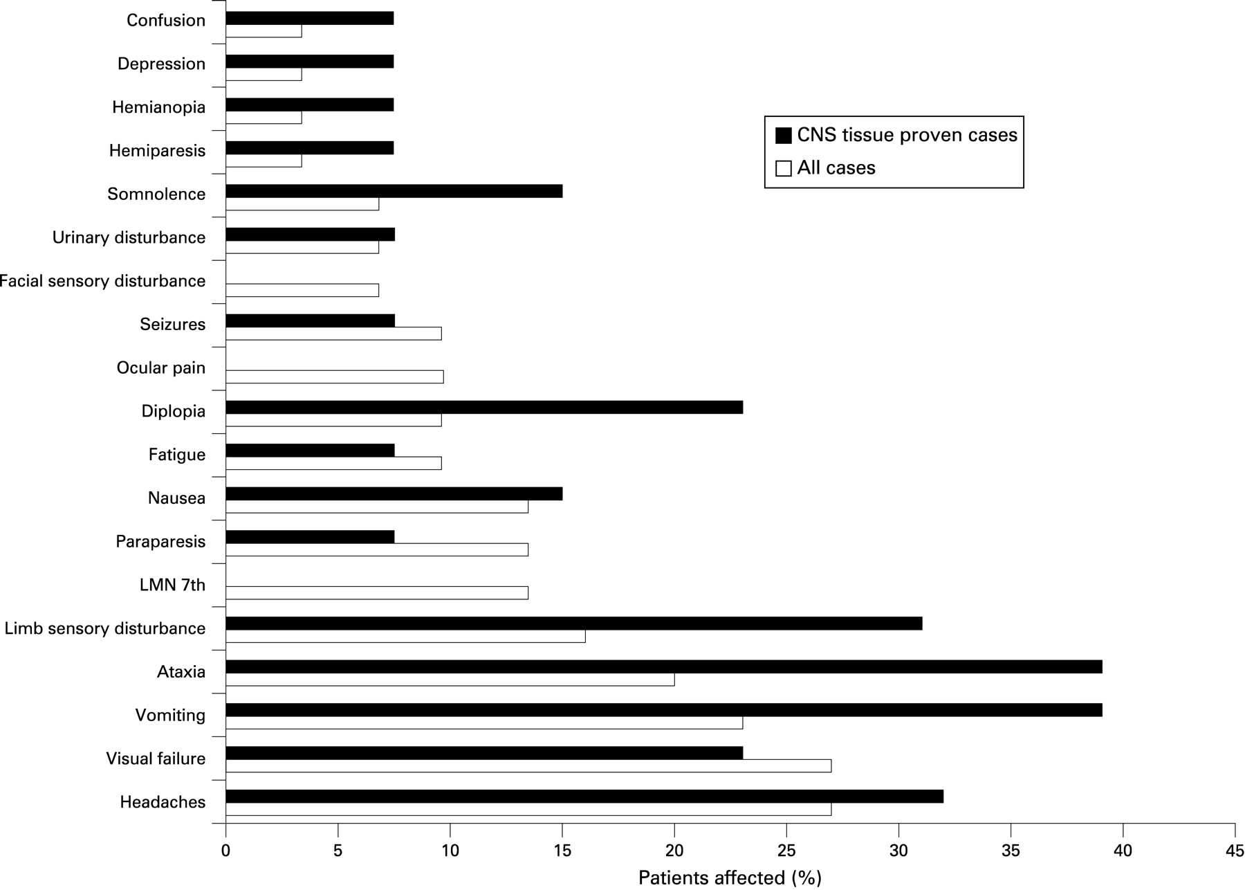
Wide-ranging neurological features were recorded—including headaches, unsteadiness/incoordination, visual failure, diplopia, fatigue, nausea and vomiting, limb sensory disturbance, seizures, memory impairment, somnolence, walking difficulty, ocular pain, facial weakness, facial sensory loss/pain, hemi-motor and visual-field loss, optic atrophy, papilloedema, monoparesis, depression, dysphasia, intention tremor, emotional lability/personality change, deafness, tinnitus and vertigo (figs 1–3).


Twenty-one (70%)/30 patients had CNS features as the first manifestation of sarcoidosis, with an almost identical proportion (9/13; 69%) in the “Definite NS” group. In order of decreasing frequency, the most common were: headaches (27%), visual failure (27%), vomiting (23%), ataxia (20%), sensory disturbance of limbs/trunk (16%), lower motor neuron facial weakness (13.5%), spastic paraparesis (13.5%), nausea (13.5%), fatigue (9.6%), diplopia (9.6%), ocular pain (9.6%), optic atrophy (9.6%), papilloedema (9.6%), seizures (9.6%), facial sensory disturbance (6.8%), urinary disturbance (6.8%), somnolence (6.8%), hemiparesis (3.4%), cauda equine syndrome (3.4%), hemianopia (3.4%), depression (3.4%) and confusion (3.4%). Optic nerve involvement was suspected in 11 (37%), with bilateral involvement in 7/11 (64%) and unilateral in 4/11 (36%). It occurred at the start of illness in five (17%). Figure 1 illustrates the relative frequency of the initial neurological features encountered for all patients, compared with those with definite NS.
INVESTIGATION FINDINGS
Blood tests
We found no consistent or typical abnormal blood tests in our study group. Serum calcium was modestly elevated in 3/30 patients. Twenty-one patients (70%) had serum angiotensin-converting enzyme (ACE) measured, and six of these (29%) were elevated. Only two out of eight patients with pure neurological presentations demonstrated significant elevation in ACE. The erythrocyte sedimentation rate (ESR) or plasma viscosity (PV) was elevated in 5/22 (23%) cases at the time of neurological presentation.
Histopathology
Kveim test
Ten of 13 patients (77%) had a positive Kveim test, of whom six also had CNS tissue confirmation. One of these cases was initially negative but became positive on subsequent repeated Kveim biopsy. Two cases were equivocal, and one was negative despite the latter having positive CNS histology.
Systemic biopsies
Eight patients had sarcoidosis supported by systemic biopsies taken from clinically affected organs. Four had positive skin biopsies; two with positive skin biopsies alone, one with positive skin and spinal cord biopsy and one with positive skin and liver biopsy. Three had respiratory tissue sampling: one had positive transbronchial lung biopsy (TBLB), Kveim test and brain biopsy, and one had positive broncheoalveolar lavage. One TBLB was negative despite positive brain biopsy.
CNS tissue
Unequivocal evidence of NS from CNS tissue was available in 13 of the 30 patients (43%). This was provided by brain biopsy in nine patients, spinal cord biopsy in two patients, and at autopsy in two cases (one of which showed both brain and spinal cord involvement).
Radiology
Chest radiographs were available in all but one patient. Fourteen of the 29 (48%) were abnormal. Of these, 13 were positive at presentation, and one patient with an initially normal radiograph was found to have abnormalities 2 years later. The most common finding was of hilar lymphadenopathy in 7/13 cases (54%), one of which had additional pulmonary abnormalities, but the remaining cases had less specific parenchymal lung changes, with one showing a pleural effusion (therefore only 7/29 or 24% had hilar adenopathy at presentation.)
CT thorax was performed in four patients who had definite chest x ray (CXR) abnormalities (hilar adenopathy, pulmonary infiltrates or fibrosis), which helped clarify the diagnosis in each case. It was also used in two patients with normal CXRs and in whom the diagnosis of neurosarcoidosis was in question, but it did not provide any further useful information.
CT brain scans were obtained on 21 patients. Nine were normal, eight showed hydrocephalus (correlating with MRI findings), three displayed enhancing lesions, and the remaining two had mass lesions. However, MRI was significantly more sensitive for other lesions, and indeed three MRI scans were abnormal while contemporaneous CT scans were unremarkable.
Cranial MRI scan results were available in 26/30 patients. Three patients who clearly satisfied the “probable” criteria for NS had normal brain imaging. The most frequently encountered abnormalities were non-specific white-matter lesions (10 patients) and hydrocephalus (eight patients). Mass lesions were seen in five patients; two involved the pituitary gland; others the cavernous sinus, basal cistern and parafalcine space (with later development of a temporal lobe mass). Significant cerebral atrophy was evident in two patients. High signal lesions in the cerebellum were seen in one case. Thirteen out of 14 patients showed meningeal or parenchymal lesion enhancement with gadolinium. Spinal MRI was performed on six patients, of which three were normal, two showed meningeal enhancement, and one had white-matter lesions.
Gallium-67 scintigraphy was available in six patients and showed characteristic uptake of radioisotope by the parotids in four of these (67%). One positive scan was accompanied by both a positive Kveim test and CNS tissue. The other positive results occurred in the absence of histological sampling. Of the two negative results, one had an equivocal Kveim reaction.
Cerebrospinal fluid
Twenty-one patients had cerebrospinal fluid (CSF) analysis. Seven had completely normal CSF constituents (despite one having a positive brain biopsy). The most consistent abnormality was an elevated CSF protein (11/21 patients, 52%) in the range of 0.56–4.3 g (mean 2 g). A CSF pleocytosis (all monocytes) occurred in nine patients (43%) with a range of 11–88 and a mean of 30 white cells. This was associated with a raised CSF protein level in six cases, but three patients had normal protein levels despite the pleocytosis. There were two cases of hypoglycorrhachia, but the CSF results of one of these with a protein elevation of 11.1 g, a low CSF glucose of 1.7 and mixed pleocytosis were not included in the analysis as they were ascribed to infection of a CSF ventriculo-peritoneal shunt. Three out of eleven (27%) patients who underwent isoelectric focusing of their CSF were found to be oligoclonal band positive (with different serum patterns, suggesting intrathecal immunoglbluin synthesis), and it was notable that all three were also found to have an elevated CSF protein. The result was equivocal in one patient (9%), and negative in the remainder.
Neurophysiology
EEG was performed in seven patients. Two were normal, one (with a known liability to seizures) showed diffuse paroxysmal activity, and four were non-specifically abnormal.
Treatment and clinical outcomes
Ninety per cent of patients were followed up for at least 18 months in this series; the mean period of follow-up was almost 10 years (115.5 months). The mainstay of drug treatment was corticosteroids, usually as a combination of oral prednisolone or dexamethasone with or without intravenous methylprednisolone. Ten patients received steroid therapy alone. Of this group, one ultimately had a severe disability, seven moderate and two minor disability. Two of the patients treated with steroids also required CSF diversion procedures for hydrocephalus. The majority showed an initially good response to treatment, but later follow-up demonstrated that symptoms often worsened when steroid doses were reduced. This usually necessitated prolonged courses of steroids or the introduction of other immunosuppressants.
Thirteen patients had azathioprine in addition to steroids. The outcome for these was: asymptomatic in one, minor disability in six, moderate disability in three and death in three patients (although one of these died of carcinoma of the colon, and the other of metastatic renal cell carcinoma). This is difficult to interpret, since the choice of azathioprine was likely to be influenced by more severe disease. The number of patients treated with any other regimen was too small to draw firm conclusions. Steroids, cyclophosphamide and hydroxychloroquine were used together in one patient who presented with hemianopia, and progressed to marked cognitive impairment, cerebellar features, dysarthria and somnolence, but with very little response, resulting in severe disability. Another who received the same combination plus methotrexate for visual failure, headaches, ataxia and facial weakness also showed little improvement with eventual moderate disability. One patient received cranial irradiation (after steroids and azathioprine had been ineffective) to control disabling trigeminal neuralgia (associated with other cranial nerve lesions, visual failure and cognitive deficits), with modest amelioration of facial pain.
Definite neurosarcoidosis
Thirteen patients fulfilled the criteria for definite neurosarcoidosis—eight males and five females (table 1). Particular attention was given to initial clinical neurological and systemic presentations, with efforts to validate the usefulness of specific investigations. Nine of these patients (69%) presented with CNS disease. Of this definite group, the most common first neurological features encountered in order of decreasing frequency were ataxia, vomiting, headaches, limb sensory disturbance, visual failure, diplopia, somnolence, seizures, fatigue, hemiparesis, hemianopia, depression and confusion.
The Kveim test was positive in 6/8 patients (75%), CXRs were abnormal in 8/13 (62%), and gallium scanning was performed in only one patient (and was positive). All had some form of abnormality on brain (12 patients) or spinal (one patient) MRI. Hydrocephalus was seen in 7/12 (58%), enhancing parenchymal lesions or meningeal enhancement in 6/12 (50%), white-matter lesions in 5/12 (42%), and pituitary-mass lesions in 2/12 (17%). The serum ACE level was elevated in 1/6 (17%). CSF protein was elevated in 4/5 (80%), cell count was raised in 3/5 (60%), and all four patients analysed for oligoclonal bands (OCB) were negative.
The final outcome was minor disability in three (23%) patients, moderate disability in five (39%) patients, severe disability in two (46%) patients and death in three (23%) patients (the cause of death at post-mortem in one patient was declared as carcinoma of the colon, despite also having clear histological features of CNS sarcoidosis from autopsy).
DISCUSSION
We have described the demographic, clinical and investigation findings and outcome of 30 new cases of NS, including one of the largest reported series of CNS tissue-proven sarcoidosis. NS has a prevalence in our study area (south-west England and South Wales) of approximately one per 100 000. A more robust figure would have necessitated a community-based survey; but we suggest that most NS cases are likely to attend secondary and tertiary referral centres. As noted by others,7 14 we found a slight male predominance—of interest since sarcoidosis preferentially affects females. Seventy per cent of our patients presented with CNS features; other case series range from 48% to 68%.5–7 10 11
The commonest NS manifestation is cranial neuropathy6 10 13 14—seen in 80% of patients in our series, and as a presenting feature in 50%. A subacute optic neuropathy occurred in over a third of patients overall and was part of the presentation of the disease in five (17%). Lower motor neuron seventh nerve palsy is a classical feature of neurosarcoidosis, unilateral in 65% and bilateral in 35%.10 We found only six patients, 23%, to have a facial palsy (all unilateral) during the course of their illness—surprisingly, not in any of the definite cases.
Of other cranial nerves, abducent nerve involvement was seen in 10% of patients (presenting feature in 7%). The well-recognised cranial nerve syndrome of the pharynx, soft palate and vocal cords from glossopharyngeal and vagus nerve involvement11 15 was seen in two cases, but neither at presentation. Cochlear nerve deafness is a recognised feature, and may even be the only feature of NS.16 Deafness occurred in two patients and was a presenting feature in one of them in our study.
Headaches were a feature at presentation in over 25% of the patients, often in a triad of headaches, vomiting and ataxia. Mass lesions were seen in five patients (involving the pituitary gland in two, and the cavernous sinus, basal cistern and parafalcine space (then temporal lobe) in the others). NS has a predilection for the base of the brain, hypothalamic and pituitary gland involvement being common, and in our study six patients had features of hypopituitarism and/or hypothalamic disease, in four as a presenting feature. One patient had isolated pituitary features, generally considered rare.17
Seizures occurred in five patients (17%), consistent with previous studies,10 14 18 and were an initial neurological feature in three (10%). Seizures in sarcoidosis are often thought to reflect serious brain pathology (mass lesions, hydrocephalus, encephalopathy or vasculopathy) and poor outcome.14 18 In our study, two patients with seizures (40%) died, but prolonged follow-up in the other three was favourable. Furthermore, in one patient who died, NS was only diagnosed at autopsy; she had never received corticosteroid or any other immune therapy. The other death was from metastatic renal carcinoma. Therefore, we suggest seizures may not necessarily imply a grave prognosis in NS.
By contrast, myelopathy in sarcoidosis is also generally thought to carry a poor prognosis,19 and none of the five such patients in our study had a benign outcome. Cauda equina involvement, considered unusual,19 20 occurred in one patient in our series (who also had CNS features), though another had typical features of a conus lesion. Initial spinal MRI in this man was normal, but repeat imaging with gadolinium showed clear enhancement, leading to biopsy of the meninges at T11 and T12, confirming the diagnosis.
Neuropsychiatric symptoms occur in NS but are generally regarded uncommon. Memory impairment was a frequent secondary neurological feature (six patients; 20%). Frank psychosis was not seen but is reported.21–23
Unfortunately, despite the almost uniquely large cohort of size of the definite group, no specific patterns or findings emerged that might help confirm or exclude NS. Almost ironically, none had facial palsy, and only a minority had systemic features.
Box 1 Proposed criteria for diagnosis of neurosarcoidosis (from Zajicek et al10)
Definite: clinical presentation suggestive of NS+presence of positive nervous system histology+exclusion of other possible diagnoses.
Probable: clinical presentation suggestive of NS+laboratory support for CNS inflammation (elevated levels of CSF protein +/or cells, the presence of OCB +/or MRI evidence compatible with NS)+exclusion of alternative diagnoses +/or evidence of systemic sarcoidosis (by positive histology, including Kveim test, +/or at least two indirect indicators from Gallium scan, chest radiograph+serum ACE).
Possible: clinical presentation suggestive of NS with exclusion of alternative diagnoses where above criteria are not met.
Zajicek et al10 formulated diagnostic criteria using investigational results such as MRI, gallium scanning, CXR, CSF and the Kveim test, to define probable and possible NS. Other authors later refined these criteria to exclude the Kveim test (rarely used because of the risk of transmitting infection), CXR and serum ACE (considered poor markers of CNS disease). Instead, they included high-resolution chest computed tomography and bronchoalveolar lavage fluid with a CD4:CD8 ratio >3.5, and a CSF CD4:CD8 ratio >5,24 although there have been no sensitivity–specificity studies for the latter two tests. These data sets spanned from 1990 to 2002, during which time period analyses of the CD4:CD8 ratios were not routinely performed, while the Kveim test was still frequently used. Our results confirmed the Kveim test as a very useful diagnostic tool for NS—a positive Kveim histology being obtained in 77% of those tested. Abandoning the Kveim is, we believe, inevitable for safety reasons but unfortunate considering its diagnostic value in an often very difficult clinical area.
In our study, 67% of patients had abnormal CSF, usually an elevated protein (in the range of 0.56–4.3 g/l) and/or lymphocytosis (mean of 30 white cells/mm3, which compares with previous work.10 Unequivocal evidence of oligoclonal bands in the CSF was found in 27% of cases (cf. 37% in Zajicek study)—neither sensitive nor specific, but perhaps helping to confirm a CNS immune process in more subtle neurological presentations. Furthermore, all samples with positive OCB in this study were accompanied by elevations of CSF protein—a finding also noted by Zajicek et al,10 which may help in the distinction from multiple sclerosis. CSF ACE measurements were not commonly performed; 3/5 samples analysed were normal, despite one having an elevated serum ACE. Two “probable” cases had CSF ACE levels above the normal reference range, in the absence of an elevated CSF protein. The utility of CSF ACE measurement in suspected or proven NS remains uncertain.25–27
Brain CT is inferior in sensitivity to MRI.28–30 In our study, white-matter lesions and hydrocephalus accounted for some 70% of all MRI abnormalities. Multiple white-matter lesions are also seen in MS, but meningeal enhancement may be distinguishing.28 Interestingly, within our definite group of patients, meningeal and/or parenchymal lesion enhancement was observed in all but one case, which may be of clinical diagnostic value.
Variable rates of CXR abnormalities are reported; the figure was 45% in our study (bilateral hilar lymphadenopathy in 25%). The “sarcoid pattern” on gallium scanning of uptake in the salivary glands or chest was seen in 4/6 patients (the reported sensitivity in systemic disease is 60–90%31). None of our patients had increased cranial uptake, as in other studies.32–34
Most treatment schedules in NS reflect experience in systemic disease. The rarity of NS, difficulty of proving the diagnosis in isolated CNS disease and its diverse presentations render randomised controlled trials difficult. Corticosteroids remain the cornerstone of treatment, but previous experience, borne out in this series, is that prolonged high doses are often needed—a good initial response often being followed by relapse or deterioration on dose reduction.10 14 35–37 Azathioprine was the commonest used second-line treatment—13 patients, with an outcome in seven (54%) of only minor disability. Too few patients received other drugs—cyclophosphamide, methotrexate or hydroxychloroquine—to draw conclusions regarding efficacy.
Acknowledgments
The authors are very grateful for the enthusiastic support and cooperation of their colleagues in Neurology and Rheumatology departments throughout south-west England and south Wales. FGJ was supported by the Neurological Research Fund established by MJ Campbell.
REFERENCES
Footnotes
Competing interests: None.
Patient consent: Obtained from the relevant Caldicott guardians.